
At 5 weeks of gestation, the transvaginal ultrasound most often shows only an anechoic lacunar image embedded in the decidualized endometrium. This isolated gestational sac, without a yolk sac or embryonic pole, nonetheless constitutes the first usable ultrasound marker. We regularly observe that its interpretation poses problems, not due to a lack of data, but because the question posed to the image (viability or location) entirely conditions the reading of the result.
Intradécidual sign and intrauterine location at 5 weeks of gestation
The first criterion to evaluate is not the size of the gestational sac but its position relative to the cavity line. The intradécidual sign, described as an asymmetrical focal thickening of the decidua surrounding the sac, allows for distinguishing a true gestational sac from a simple intra-cavitary fluid retention.
At this stage, the gestational sac provides information about location, not viability. A properly implanted intrauterine sac excludes, in the vast majority of cases, an ectopic pregnancy, which remains the clinical priority at 5 weeks of gestation. The absence of an embryo or a heartbeat is physiological and should in no way lead to a diagnosis of a non-viable pregnancy.
We recommend systematically documenting the position of the sac in relation to the cervical canal and the uterine horns. A low or angular implantation, sometimes difficult to differentiate from a cornual pregnancy, significantly alters the management approach.
The angular pregnancy, implanted at the junction of the uterine horn and the cavity, represents a diagnostic pitfall that popular articles rarely address. To better understand what to see on ultrasound at 5 weeks of gestation, the distinction between location and viability remains the starting point.
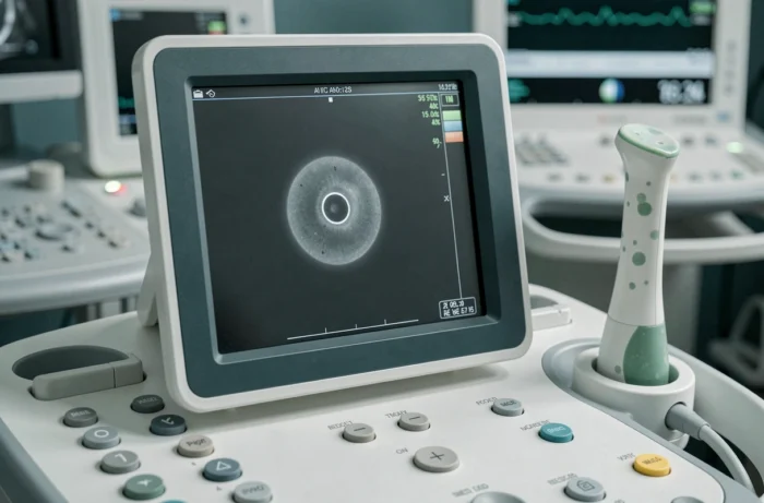
Measurement of the gestational sac: mean diameter and correlation with hCG levels
The mean sac diameter (MSD) is calculated by averaging three orthogonal measurements. This value allows for estimating gestational age with a margin of error that remains wide at this term.
A visible sac on transvaginal ultrasound generally corresponds to an hCG level exceeding the discriminatory zone. Below this threshold, the absence of an intrauterine sac does not allow for a conclusion and necessitates close biological monitoring to assess the doubling kinetics of serum hCG.
The correlation between MSD and hCG is not linear. A small sac with a high hCG level may suggest a pregnancy of uncertain viability, while a sac of size consistent with a normally progressing hCG indicates a normally evolving pregnancy. This MSD/hCG ratio constitutes a more reliable triage tool than each parameter taken in isolation.
Common measurement pitfalls in early ultrasound
- A retroverted uterus alters the quality of the transvaginal image and may lead to underestimating the MSD, especially if the probe is not oriented along the axis of the uterine body.
- The presence of a pseudo-sac (intra-cavitary fluid collection mimicking a gestational sac) remains possible, particularly in ectopic pregnancies secreting hCG. The absence of a hyper-echoic trophoblastic ring (double decidual sign) points towards this differential diagnosis.
- An overly full or empty bladder alters the uterine position and the acoustic window, generating measurement artifacts that distort the estimation of the MSD.
Minimum delay before diagnosing a non-evolving pregnancy on ultrasound
No diagnosis of a non-evolving pregnancy should be made based on a single ultrasound at 5 weeks of gestation. Recent recommendations emphasize a minimum re-evaluation period before any definitive conclusion. A gestational sac without a visible embryo requires a follow-up ultrasound, generally one to two weeks later.
The criterion retained is based on the MSD. As long as the mean diameter of the sac remains below the critical threshold (established by the scientific societies of radiology and gynecology), the absence of an embryonic pole cannot be interpreted as a clear egg. This threshold has been raised in recent years specifically to avoid falsely positive diagnoses of non-evolving pregnancy.
Management between two follow-up ultrasounds
Biological monitoring through serial hCG measurements (every 48 hours) allows for documenting hormonal kinetics. A regular increase in hCG constitutes a favorable indicator, even when the ultrasound image remains poor. Conversely, stagnation or a drop in levels points towards a non-evolving pregnancy or an ectopic location.
We emphasize the necessity of correlating the ultrasound with the complete clinical context:
- Presumed date of ovulation and cycle regularity: late ovulation shifts the entire ultrasound schedule and explains a smaller than expected sac.
- Associated symptoms: lateral pelvic pain or heavy bleeding justify an earlier re-evaluation, without waiting for the standard delay.
- History of ectopic pregnancy or early miscarriage: these elements modify the vigilance threshold and the frequency of monitoring.

Yolk sac and embryo: chronological markers in early ultrasound
The yolk sac usually appears before the embryonic pole. Its absence at 5 weeks of gestation is not a warning sign if the gestational sac is small and intrauterine. The yolk sac becomes expected when the MSD exceeds a certain threshold, which varies according to the protocols of each center.
The embryonic pole, on the other hand, is generally identifiable only from the end of the fifth week of amenorrhea, often closer to 6 weeks of gestation. Cardiac activity follows shortly after the appearance of the pole. Requiring these structures at 5 weeks of gestation amounts to applying an inappropriate reading grid for the term, generating unnecessary concerns and sometimes premature decisions.
The ultrasound at 5 weeks of gestation remains a landmark examination. Its value lies in confirming an intrauterine pregnancy and establishing a baseline for subsequent follow-up. Any extrapolation on prognosis based on a single early image constitutes a methodological error, regardless of the imaging center’s level of equipment.